
Concurrent fNIRS and TMS
Concurrent fNIRS and Transcranial Magnetic Stimulation (TMS) allows for direct investigation of cortical brain activation and connectivity. Moreover, the effects of repetitive TMS (rTMS) in distinct brain regions without quantifiable behavioral changes can be objectively measured through fNIRS.
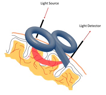
Since fNIRS does not use electrical (nor magnetic) fields, it is suitable to be combined with TMS without any particular technical precautions. Furthermore, as fNIRS is an optical signal, the strong magnetic fields generated during TMS do not cause artifacts or interference in the fNIRS data. The TMS coil may therefore be placed directly over fNIRS probes, making it possible to measure hemodynamic changes directly under the stimulated site.
fNIRS: A Technique for Neuroimaging During TMS - Promising fNIRS-TMS Applications
Compared to many modalities, fNIRS is still relatively new. More studies are needed in the field of concurrent fNIRS and TMS, but all indications suggest that this combination would be beneficial for a variety of applications:
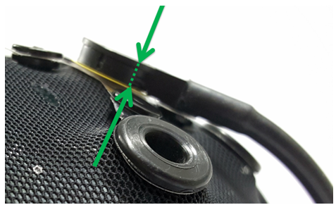
Top: NIRx’s Low-Profile Probes: Easy to Set Up and Stable. Steady probe placement is vital for high-quality fNIRS data.
In vivo studies of the effects of single pulse TMS and rTMS. rTMS is used for treatment of many psychiatric disorders. fNIRS seems to be the only possibility for in vivo assessment of cortical activation patterns during rTMS treatment sessions. Furthermore, fNIRS is being evaluated as a potential biomarker for the assessment of the outcome of rTMS treatments or as a predictor of their outcome.
TMS guided fNIRS imaging. The limited spatial specificity of fNIRS could be overcome with the aid of targeted TMS mapping, making the fNIRS signal more specific to the functional cortex. The optimal location of the probes can be identified by means of single pulse TMS.
fNIRS guided TMS stimulation. Cortical activation patterns identified by means of hemodynamic changes can be explored as TMS stimulation sites.
Advantages and Considerations for fNIRS-TMS studies
Combining fNIRS with TMS has several advantages, including: the absence of electromagnetic interference between the TMS pulse and the optical signal, better temporal resolution than fMRI or PET, and both a small and portable imaging unit. However, several technical considerations are important:
The overall height profile of the optodes needs to be minimized to prevent magnetic field loss due to increased distance between the coil and the scalp. NIRx provides optodes specially designed for TMS, with an overall height profile less than 10mm.
To streamline data analysis, TMS pulses must be synchronized with fNIRS data acquisition. All NIRx devices have either 4- or 8-bit digital trigger inputs which can be connected to the trigger output ports of TMS devices.
The mechanical contact between the TMS coil and the optodes can perturb the optical contact between the optode tip and the skin, possibly causing artifacts. NIRx provides a complete TMS solution, including optode stabilizers and retaining caps, that make the complete setup much more robust.
TMS stimulation might produce hemodynamic changes not only through neurovascular coupling but through other vascular mechanisms that need to be accounted for when analyzing fNIRS traces. The NIRx Support Team provides comprehensive scientific support throughout your research projects starting from experiment planning to data analysis.


Top Left: The NIRx NIRScap with integrated TMS optodes. The optodes are completely compatible with TMS stimulation. Their low profile allows for very close placement of the TMS coil.
Top Right: The NIRx TMS-compatible overcap, placed over the NIRScap. The retaining cap stabilizes the setup making it more robust to TMS coil contact artifacts and motion artifacts in general.