

Click to Enlarge
Quality Data without Compromise
Built-in features such as automatic calibration and diagnostics are put to work to assure that your experiment begins by collecting the best data possible. The NIRStar signal quality indicator allows you to effortlessly review the integrity of any incoming data.

Click to Enlarge
Adapted to Your Needs
Easily move between common, predefined montages (from a set that allows the user to have a quick setup of the hardware configuration according to the desired experiment design) or customize your own and save them for use in future measurements.
Click to Enlarge
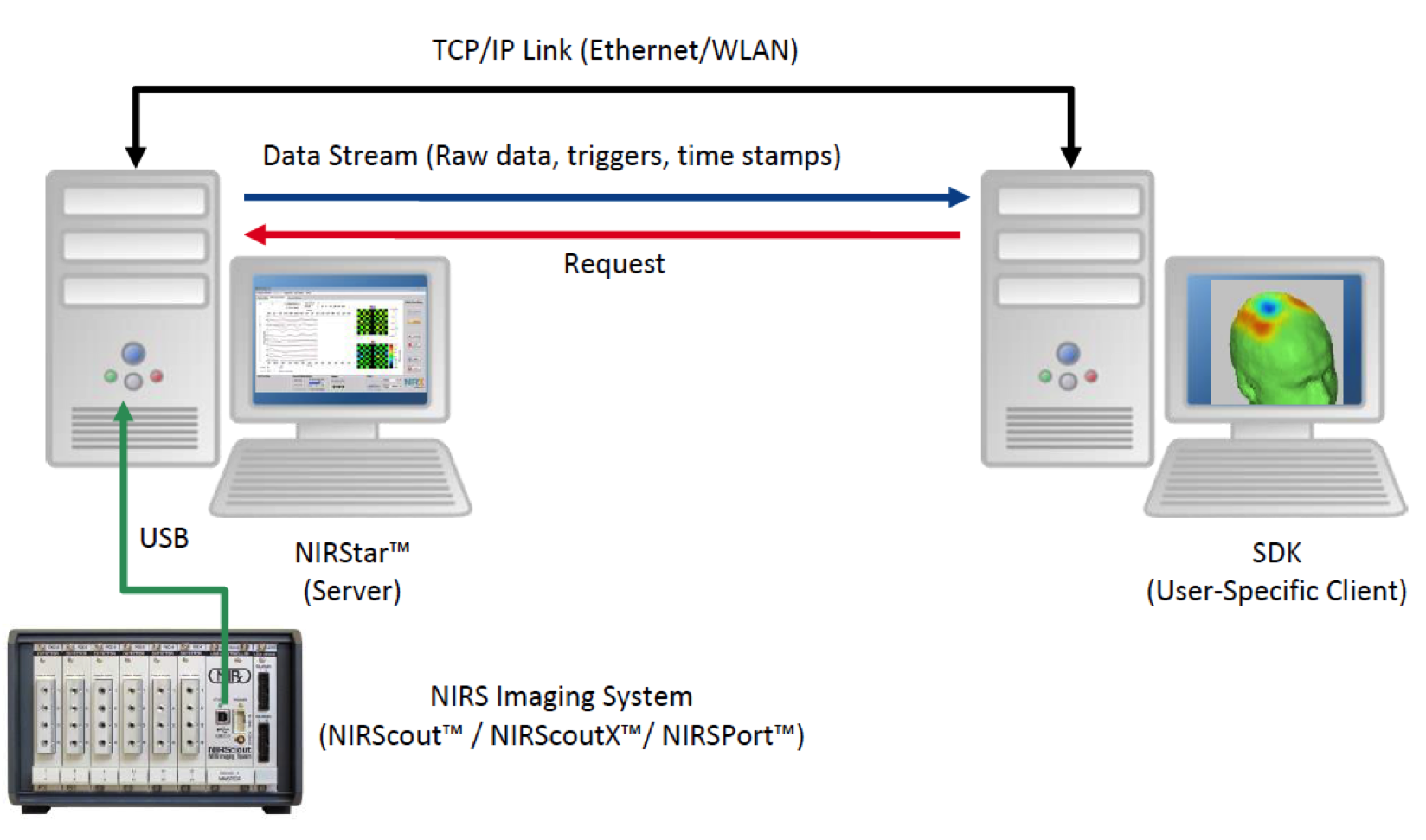
Interact with Your Data
Out of the box, NIRStar is capable of delivering realtime block averages and activation views allowing clear subject monitoring and the ability to compare events while recording.
Streaming of data to another PC allows you to process the data in real-time enabling the use of machine learning algorithms for multi-modal BCI with NIRS and EEG.

Click to Enlarge
Solid Results with NIRx Flexibility
NIRStar 14.1 allows the user to automatically export recorded datasets to the Homer2 format. To the left is an example of Hyperscanning data - NIRStar automatically separates each subject’s data into its respective folders.
Additional features of nirstar include:
• Automated instrument recognition upon software startup.
• Ability to run full or partial sensing configurations.
• Tandem operation for all instrument types.
• Programmable source control to support multisite brain studies (e.g., left and right hemispheres).
• Automated logging of input triggers from 3rd party devices.
• Programmable specification of output triggers to 3rdparty devices (NIRScoutX only).
• User-controlled manual trigger input.
• Multiple real-time display screens.
• Montage viewing options: Glass view, optode locations and standard EEG positions (international 10-10 system).
• Sensor registration onto anatomical displays (scalp, cortical, MNI brain).
• Real-time updates of hemodynamic response to event related protocols.
• Real-time hyperscanning topographic displays (Hbdeoxy, Hboxy, Hbtotal).
• Programmable display capability to highlight preselected sensor locations.
• Automated gain adjustment to maximize dynamic range of measurement.
• On-line frequency filtering of displayed results.
• User GUI for specification of custom optode configurations.
• Automated helpful suggestions to facilitate system use.
• Real-time system status reporting display.
• User directed comment box for information logging.