
Other fNIRS Applications
fNIRS Neuroimaging
Disturbances in brain function can have immediate, severe and lifelong consequences. Monitoring activity of the brain holds considerable significance to a broad range of clinical fields (e.g., neonatology, psychiatry, gerontology, emergency care medicine, neurology) and to a variety of practical problems that range from the study of learning disorders to issues of Homeland Security. Underscoring the expected utility of fNIRS to this area is the well-founded idea that accompanying neural activation is a vascular response that serves to replenish activated tissue with improved blood flow. Functional studies have shown that the brain activation produces a spatially distributed and temporally varying response. Techniques such as electroencephalography (EEG) and its magnetic equivalent, magnetoencephalography (MEG), provide a high degree of temporal resolution but have markedly reduced spatial resolution, especially for EEG, compared to anatomic imaging techniques (e.g., MR, CT). An imaging modality that is proving to have significant impact in investigative studies is functional MRI (fMRI). This technique is sensitive to the vascular response resulting from neuroactivation, specifically to the level of deoxyhemoglobin. While the utility of fMRI continues to expand, it is also clear that the technique has a number of limitations that are not encountered using near infrared techniques.
Imaging funcational Response of the brain
GLM image of fingertapping response
The ability of near infrared light to penetrate through the cranium and interact with the cerebral cortex has prompted investigators for many years to explore the utility of near infrared methods. NIRx fNIRS imaging systems extend NIR spectroscopic studies to provide for collection of 3-D tomographic images that detail the dynamics of the vascular response to various forms of neuroactivation. Unlike in functional Magnetic Resonance Imaging (fMRI) the optical method can distinguish between changes in deoxyhemoglobin levels caused by a variation in blood volume or blood oxygenation. In addition, optical methods allow for experimental paradigms that are highly impractical or infeasible with other functional imaging modalities due to restricted patient access and limited freedom of patient movement. While studies using fNIRS for neuroimaging applications are now beginning to gain momentum, the current literature from the research community has made it evident that the application opportunities in the clinical, research and commercial markets are vast.
An example of the image quality obtained using the NIRScout imager is shown in figure 8. Here we imaged the oxyhemoglobin response to activation of the motor cortex in response to finger tapping. The image shown is a result of an GLM computation using a boxcar model function applied to a time series of unconstrained reconstructed 3D images overlaid on a representative brain map obtained by MR. The inset shows an orthogonal view of the image revealing that the region of activation is truly subsurface.
Value/Advantage
High Temporal Resolution
Excellent intrinsic sensitivity to hemoglobin
3D imaging capability
Measurements possibly with a variety of tasks
Easily adapted to incorporate other measurement techniques (EEG, fMRI)
Economical, portable, scalable
NIRS Breast Imaging
Breast cancer is a principal concern in women's health. Currently, one woman in eight will develop breast cancer at some time during her life. Clinical experience has shown that early detection is key to reduced morbidity and long term survival. Presently, x-ray mammography is the gold standard used for routine screening and monitoring the response to therapy. Many women find the experience of a mammogram, with its need for breast compression, distinctively unpleasant. In addition, the reliance of x-ray findings on anatomical features that are present only in the more developed tumors presents a notable challenge for early detection. Still other limitations of the technique include is its limited use in younger women having radio-dense breasts and the reliance on use of ionizing radiation.
Breast Cancer
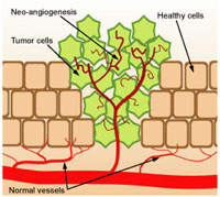
Tumor neo-angiogenesis
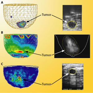
A hallmark in the development of a cancerous tumor is its ability to trigger the formation of new blood vessels to provide nutrients necessary for its growth. This process known as neo-angiogenesis does not follow the normal angiogenic mechanisms found in healthy tissue, and results in the development of a vascular network that is largely devoid of the normal control mechanisms and has vessels that exhibit aberrant morphology and impaired perfusion. Frequently this produces disturbances in intercellular fluid balances that can add to diffusion barriers caused by the distorted vascular architecture. A consequence of this is that tumors are thought to function on the brink of hypoxemia. Clinically this is known to interfere with the delivery of chemotherapeutic agents and response to radiotherapy. It is our view that the tumor environment, with its impaired perfusion, increased blood volume resulting from increased vessel density and enhanced levels of deoxyhemoglobin, is ideally matched to the expected sensitivity of NIRx systems to identify discriminatory features linked to tumor angiogenesis and other factors associated with tumor growth. Notably the expression of these features can be expected to vary in accordance with different forms of functional provocations. For instance, it is logical to expect that the existence of an impaired vascular network will be more susceptible to induced hypoxemia than will be the surrounding tissue. Similarly, maneuvers that produce changes in the bulk perfusion of the breast can likewise be expected to reveal local disturbances in flow associated with the developing tumor. Still other types of functional disturbances might be observable as a consequence of an aberrant vascular network. One example is the idea that developing tumors might cause regional disturbances in the normal control mechanisms that modulate blood flow to tissue. Clearly the implication is that there is a host of vascular dynamic behaviors that can be exploited to act as a contrast feature to detect neo-angiogenetic vascular disturbances. These include vascular dynamics at rest as well as responses to a variety of possible external stimuli (e.g., breathing challenges, temperature changes, etc.). Shown here are a few exemplary results that support this idea obtained using the DYNOT 232 imager on subjects diagnosed with breast cancer.
The image in panel A was produced from a subject asked to undergo a prolonged breath hold (60 sec) in an effort to induce hypoxemia. The image shows a high contrast feature positioned in the anterior region of the breast near the nipple whose size and location coincides well with comparative findings from ultrasound.
In panel B we show a result obtained wherein blood flow to the breast was affected by deep regulated breathing. The expected finding here is that, in healthy tissue, changes in blood flow caused by deep breathing will be uniformly synchronized throughout the breast. A map of time delays in the response of the venous vasculature to the induced respiratory rhythm (phase map at the respiratory frequency) can reveal this. In the healthy breast we see that the response is largely uniform. In the tumor-bearing breast, we see a large phase contrast feature spanning from the lower medial to the upper lateral quadrant indicating the occurrence of a time delay in venous return in response deep breathing. Significantly, this feature agrees well with the presence of a large tumor revealed by x-ray having a similar location.
In panel C we have explored yet another well-known feature of vascular physiology - the modulation of the natural vascular rhythms. This phenomenology occurs in response to complex interactions involving both local tissue and central control mechanisms (e.g., neural modulation of the microvasculature) that serve to redirect blood flow on a local level. This behavior is readily observable from a time-frequency analysis of images obtained from the breast of a subject at rest. In the example shown we present a contrast map that reveals the presence of a diffuse region having altered modulation. Comparison of this map to the corresponding map obtained using ultrasound indicates that the region of tissue exhibiting an altered response extends well beyond the boarder of the tumor. This finding raises the intriguing possibility of early tumor detection (perhaps sizes well below that seen by x-ray) based on the response of the surrounding tissue to the growing tumor.
Significantly, the examples cited represent distinctly different contrast mechanisms. Common to all is the fact that the observed features are associated with functional disturbances. It is our view that many more such features can be defined and that these will serve to discriminate the presence of cancerous tumors and differentiate them from other pathologies. It deserves emphasis that in all cases shown, results were obtained without breast compression, need for injectable contrast agents or use of ionizing radiation. On a fundamental level, we believe that our technology is intrinsically better suited for early detection of tumors than other techniques because there is a better match between features of tumor biology and the type of sensing methods used to explore these.
value/advantages
Intrinsically greater sensitivity to early detection of tumors
No compression required; increased patient comfort
No injection required
Added information gathered from simultaneous bilateral scans
No interference from dense breasts or implants
High patient throughput
No ionization radiation; hense, more frequent screening without corresponding health concerns
Peripheral Vascular Diseases
Many believe that the growing problem of obesity will significantly expand the incidence of Type II diabetes, with an ensuing increase in peripheral vascular disease. Even in well-controlled diabetics, long-term damage to the peripheral microvascular bed is evident. We believe that the availability of compact, robust devices that can be worn comfortably for extended periods of time could prove key in gaining basic insight into early microvascular changes. Such measures could also provide objective feedback of the influence of lifestyle changes and the long-term effects of drugs aimed at stabilizing carbohydrate disorders.
Investigating PVD with DYNOT
Optical Imaging of PVD
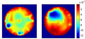
Protocols for forearm provocation involving long-term diabetics have produced results revealing a statistically significant inverse relationship between the rates of recovery following venous occlusion and plasma hemoglobin A1c levels, an established marker of long-term control of glucose metabolism. In addition, reduced recovery rates often are accompanied by a marked decrease in vascular contrast features seen in cross section suggestion the presence of occlusive disease (see figure to the right, left panel: healthy; right panel: PVD patient).
Studies have furthermore demonstrated clear relationships between recovery rates and factors that influence peripheral vascular resistance (PVR). The assessment of this parameter holds high significance in acute care medicine. Currently PVR measures require placement of a Swan-Ganz catheter in the pulmonary artery, which is a highly invasive procedure not without risk.
Other areas where DYNOT measures could prove particularly useful are in the assessment of compartment syndrome and for detecting focal occlusive events associated with sickle cell crises. The latter is a disease that mainly effects children and presents clinicians with few options for determining when emergency transfusions are warranted. With DYNOT, assessment of perfusion disorders can be performed without the risk of anesthesia that is inherent to MR studies of young children.
Value/Advantages
Excellent sensitivity to perfusion state of microvascular bed
Feasibility of employing sensor arrays suitable for long-term monitoring
Economical, compact systems
Competing Technologies
Vascular ultrasound, x-ray angiography, and MR-angiography are the main technologies employed to assess blood flow disorders. All are costly, and none is well suited to explore that functional state of the microvasculature.
Small Animal NIRS Imaging
Noninvasive longitudinal investigations of laboratory animals are increasingly gaining favor among the biotechnology and pharmaceutical industries. The information derived is often far more relevant than can be gained from acute terminal studies, and in many instances the number of animals and associated costs is significantly reduced. In recent years optical studies with live animals has gained favor as a tool to explore molecular expression. Among the molecular imaging methods being employed, reporter gene studies with Luciferin or Green Fluorescent Protein are now widely used. While effective, this approach does not lend itself to translation to human trials. On the other hand, measurements based on the hemoglobin signal are fully translatable. We believe that, in many instances, the hemoglobin signal can be used as a surrogate marker for factors that ultimately impact on cell metabolism. Another argument supporting use of fNIRS in animal studies is its ability to directly assess whether candidate drugs have desirable or undesirable effects on blood flow. Given the central role of hemoglobin to body function and the integrated nature of vascular reactivity, the merits of this measure hold whether or not the considered drug is vasoactive.
Small Animal Imaging
Rat NIRS imaging software
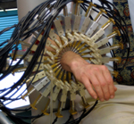
A four-wavelength small animal imager is available that can be easily adapted to explore various target tissues in mice, rats and other small animals. Notably the particular measuring head developed for rat brain studies is MR compatible.
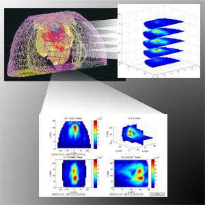
A photograph of the measuring head is shown above. The unit features a three point stereotactic holder with optical fibers being introduced into the mouth, ears and from the top of the head. The top array is effectively spring loaded to ensure good tissue contact and has two axes of motion. An example of the 3D image quality achieved of the rat head and new image display capabilities is shown to the right. Seen is volume rendered image based on a 7-tissue segmented finite element mesh of the rat head depicting the response to a 3 second forepaw stimulation at 10 Hz. New image display capabilities include an interactive MATLAB based 3-axis orthogonal views, user directed exploded views, among other features.
Value/Advantages
fNIRS offers a low cost solution to molecular imaging and evaluating of effects of investigative drugs. Currently available are custom configured and MR compatible measuring heads for brain and abdominal investigations that are suitable for animals ranging in size from mice to guinea pigs.