Our Story
fNIRS and NIRx
New Technologies Require New Understandings:
A Collision of Ideas Born in Brooklyn
As often happens in science, new insights are drawn from the most unlikely of interactions. In our case, it involved common understandings from the fields of Life Sciences and Nuclear Physics: the problem of scattering. Whether the form of energy is light or a neutron, the physics of scattering is the same. Armed with this understanding, in 1988, Drs. Randall Barbour from the SUNY Downstate Medical Center and Raphael Aronson from the now NYU Polytechnic University were the first to show that whereas individual scattered photons have an unpredictable path, bulk movements can be defined [1]. Using mathematical transforms that leveraged this understanding, these investigators produced the first 3D images of otherwise obscured features from an opaque medium. The result was the birth of a field of imaging that today is opening new vistas in our understanding of tissue function: optical tomography.

1) Individual photons traveling through a scattering medium

2) The characteristic ‘photon banana’ of bulk photon movement

3) Multi-distance source-detector configuration (tomography) to identify cortical activation

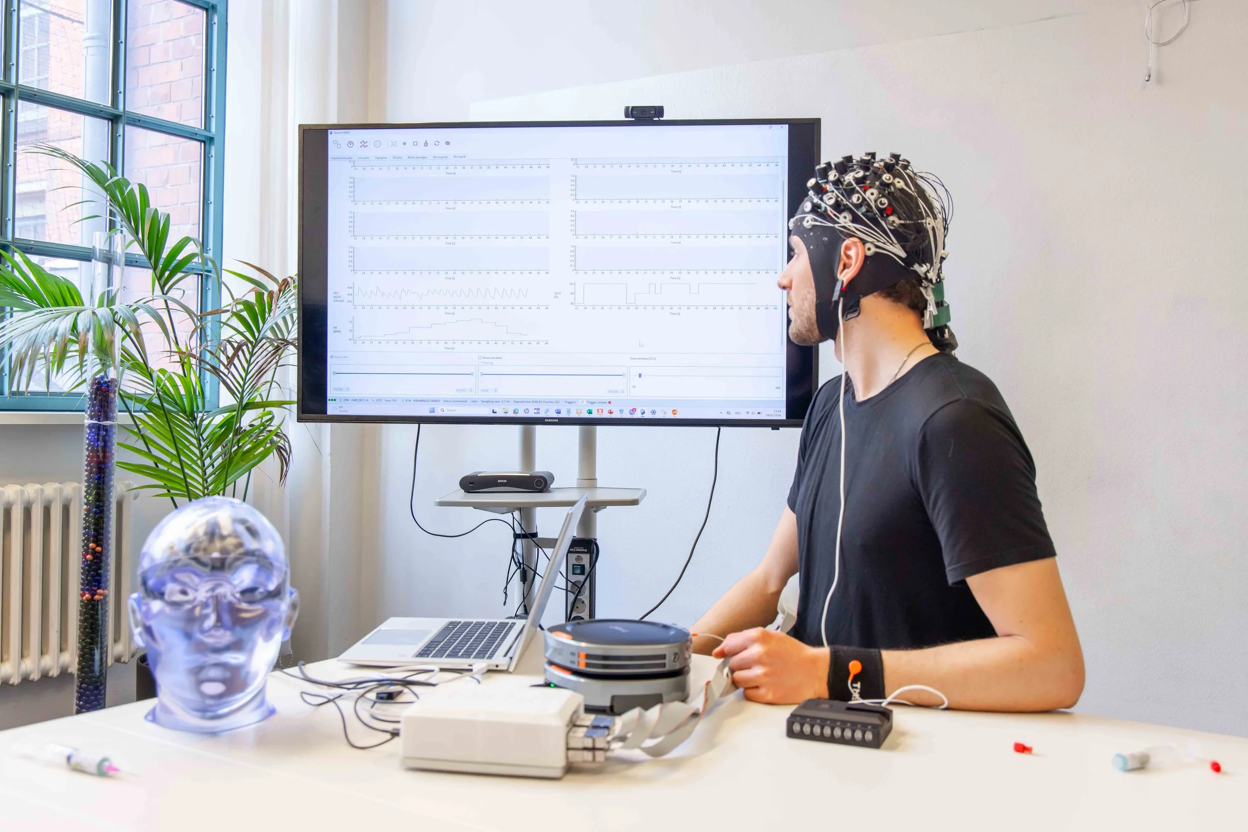
4) Tomographic brain imaging with fNIRS system by NIRx
The fNIRS Signal

A simple demonstration that red light is shines through tissue.
Accompanying neuroactivation, is a coupled hemodynamic response that is sensitive to a host of features of coordinated brain function. Relating these measures to the seemingly endless breadth of human behavior is a principal aim of many scientific investigations.
Learning, language acquisition, sensory and motor functions, emotion, social interactions, and the influence of a host of disease processes all can be explored from measures of the fNIRS signal. Most important, the resources needed to accomplish such measures are easily made wearable, resistant to motion artifacts, economical and can be performed in the natural environment. Further, employing harmless light sources, NIRS is safe for studies with infants and adults as well.
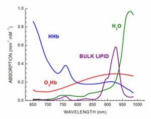


In contrast to information derived from the fMRI BOLD signal that can identify portions of the hemodynamic response, the fNIRS signal offers added information regarding the coupling between tissue metabolic activity and its blood supply. Supporting direct measures of both oxy- and deoxyhemoglobin, with deep tissue penetration, the NIRS signal supports real-time evaluation of related biometrics that are known to influence brain function.
The NIRx Team:
From First Images to Comprehensive Integrated Solutions
Twelve years after Dr. Barbour and Dr. Aronson’s discovery, the group formed NIRx to focus and promote the group’s instrumentation and software advancements. Today NIRx offers a host of integrated technology solutions that support a wide range of investigative aims. Whether the goal is to explore early language acquisition in infants [2], motor movements in the natural environment [3], BCI applications [4] or new understandings involving coordinated actions between sensory systems [5], NIRS imaging solutions from NIRx constitute a comprehensive resource that meet the most demanding applications.
NIRx Today:
Versatile fNIRS Solutions and Dedicated Scientific Support
Our innovations in head gear design provide for fast setup and hours of comfortable wearing. The many real-time display and 3rd party device control capabilities make our NIRStar system control software the most advanced and user friendly in the industry. Supporting all of our platform configurations, including tandem system operation and hyperscanning, NIRStar offers unmatched performance. Complementing this is our new NIRStim experimental design software that offers a host of innovative capabilities that are supportive of social interaction studies. Further adding to this is our growing suite of data analysis resources (nirsLAB, NAVI) that we provide as a courtesy to the scientific community.

All of these capabilities are accessible by custom configurable scalable hardware resources that are further adaptable to integration with any number of modalities, such as: EEG, fMRI, and neuromodulation technologies including tDCS and TMS. In all, NIRx offers the most comprehensive range of solutions that are backed by a team of dedicated developers, scientists and support staff that have a proven track record of sustained innovation whose focus is purposefully restricted to NIRS solutions. We adopt this approach because of our unwavering belief that NIRS offers the largest range of solutions among all alternative forms of neuroimaging.


Have an Idea?
Wish to Submit a Grant Proposal?
NIRx developers and scientists are among the most experienced in the industry. NIRx often collaborates with investigative teams in the preparation of grant proposals. The NIRx scientific team has published nearly 300 peer review and conference reports and has been continuously funded by granting agencies in the US and Europe since 1993.
For detailed information on custom solutions: fNIRS and instrument specifications: NIRx contact page

Have a need for a custom configured resource?
Whether it involves new innovation in hardware design, custom software development or integration with 3rd party resources, NIRx frequently provides custom design resources to meet the most demanding applications.
For detailed information on custom solutions: fNIRS and instrument specifications: NIRx contact page
The Expanding Group of NIRx Users
Today NIRx systems are used by thousands of customers in distinguished research institutions. This growth has been accomplished through a steadfast commitment to excellence in system design, reliability and advanced product support.
References
[1] R.L. Barbour, J. Lubowsky and R. Aronson, “Method of Imaging a Random Medium”, US Patent no 5,137,355, filed June 8, 1988, awarded August 11, 1992.
[2] J. Gervain, “Plasticity in early language acquisition; the effects of prenatal and early childhood experience”. Current opinion in neurobiology 35, 13-20 (2015).
[3] S.K. Piper, A. Krueger, S.P. Koch, J. Mehnert, C. Habermehl, J. Steinbrink, H. Obrig, and *C.H. Schmitz, “A wearable multi-channel fNIRS system for brain imaging in freely moving subjects,” Neuroimage, doi:pii: S1053-8119(13)00700-3. 10.1016/j.neuroimage.2013.06.062 (2013).
[4] Lee, Min-Ho, Siamac Fazli, Jan Mehnert, and Seong-Whan Lee. "Subject-dependent classification for robust idle state detection using multi-modal neuroimaging and data-fusion techniques in BCI." Pattern Recognition 48, no. 8 (2015): 2725-2737.
[5] Ling-Chia Chen, P. Sandmann, J.D. Thorne, C.S. Herrmann, S. Debener. "Association of Concurrent fNIRS and EEG Signatures in Response to Auditory and Visual Stimuli." Brain topography: 1-16 (2015).