
Beyond the Brain: Physiological Signals in fNIRS Research
Functional Near Infrared Spectroscopy (fNIRS) is a flexible and non-invasive imaging technique, making it particularly attractive for studies in naturalistic, mobile, and social environments. One of the major advantages of fNIRS is the ease with which it can be combined with other modalities such as peripheral physiology. Indeed, recent progress in fNIRS methods has identified two ways that physiological data can be incorporated into fNIRS analyses.
First, fNIRS is designed to measure the cerebral hemodynamic responses evoked by neural activity. However, as the technique measures light absorption through both cerebral and extracerebral tissues, systemic physiological processes, such as cardiac pulsation, respiration, and autonomic fluctuations, can significantly contaminate the signal (Figure 1). Addressing these confounds is therefore critical for accurate interpretation.

Figure 1: A graphic of the physiological signals reaching the detector and their origins. Functional brain activity comes from the cerebral cortex, but respiration, cardiac, and Mayer wave data reaching the detector come from both the cerebral and extracerebral layers. Sensors measuring systemic signals in the body, such as EMG, respiration, and short-distance detectors, can help remove those artefacts.
The second application of combined fNIRS-peripheral physiology integrates both in order to investigate the brain-body relationships by examining how peripheral physiology interacts with neural responses (joint analysis).
This dual motivation, cleaner data and richer insights, has led to growing interest in what is now termed Systemic Physiology-Augmented fNIRS (SPA-fNIRS) (Scholkmann et al., 2022). The following sections discuss the nature of these physiological confounds, the methods used to address them, examples of SPA-fNIRS in recent publications, and how the NIRxWINGS2 system facilitates both robust denoising and advanced multimodal research paradigms.
SPA-fNIRS for Denoising
fNIRS captures light absorption through tissue, and that means it also picks up non-neural signals from the scalp, skin, and superficial vasculature. As Tachtsidis and Scholkmann (2016) explain, these systemic influences can introduce false positives or negatives, distorting experimental results (Figure 2). Even subtle changes in heart rate variability or respiration can shift the fNIRS baseline, mimicking brain activation or obscuring it entirely.

Figure 2: Visualization of the six components of the fNIRS signals. (a) An incorrect assumption is that the fNIRS signal represents only changes associated with functional brain activity due to neurovascular coupling. (b) In reality, the fNIRS signals comprise six components, so that five components are potentially confounders in every fNIRS study. The contribution of the components to the fNIRS signal is visualized by color-coding (red: 100%, white: 0%). As the figure shows, four of these five potential confounds relate to systemic signals of the sort measured by the NIRxWINGS2. From Tachtsidis and Scholkmann (2016).
Peripheral Signals vs Short Channels
To combat this issue, physiological signals can be measured concurrently and used to model and regress out non-neural variance (Figure 3). In fNIRS, there are two primary approaches to measuring systemic influences: short-distance detectors capture extracerebral systemic signals (see our webpage on short distance detectors) but by definition do not measure from the cerebral compartment, while dedicated peripheral physiology sensors directly measure specific physiological modalities, potentially allowing for the removal of cerebral and extracerebral systemic signals. Indeed, these approaches are complementary and can in principle be combined. This integration leads to cleaner fNIRS data.
As von Lühmann et al. (2020) demonstrated using advanced signal separation methods, adding peripheral signals alongside short channels led to improvements of 12–16% across a set of data quality metrics. Likewise, Gugliemini et al. (2025) showed that the impact of changes in breathing was not fully captured by short-channel data, but could be accounted for with additional peripheral signals.
However, collecting and synchronizing these signals has historically been a technical hurdle.
NIRxWINGS2: Seamless Systemic Physiology Integration
NIRxWINGS2 is NIRx Medical Technologies’ dedicated module for capturing peripheral physiological data alongside fNIRS. Designed as an extension for the NIRSport2 system, NIRxWINGS2 empowers researchers to conduct SPA-fNIRS with minimal setup complexity.

Figure 3: NIRxWINGS2 wearer equipped with respiration belt, bipolar EMG sensors, PPG, GSR, temperature sensor, and two NIRSport2s with 32x32 source-detector montage. (NIRx, 2025)
What NIRxWINGS2 Measures
Respiration – Chest expansion and breathing rhythm
Electrodermal Activity (EDA/GSR) – Sympathetic arousal
Skin Temperature – Peripheral vasodilation/constriction
Pulse Oximetry (PPG) – Heart rate and SpO₂
Bipolar signals such as EMG and ECG – Muscle and cardiac activity
All data streams—including triggers— are fully synchronized within the NIRx Aurora software, eliminating the traditional headaches of integrating multiple systems. Wireless data transmission and onboard storage allow participants to move freely, even outside the lab, while still capturing high-quality multimodal datasets.
SPA-fNIRS Enriches fNIRS Experiments

Figure 4: Participant in the Li et al. 2024 study on developing a human-machine trust evaluation method for High-Speed Train Drivers
Recent research demonstrates the power of Systemic Physiology-Augmented fNIRS (SPA-fNIRS) to capture complex interactions between brain activity and peripheral physiological signals.
Examples of Recent SPA-fNIRS Research
Li et al. (2024) developed a trust evaluation method for high-speed train drivers using multimodal physiological signals to enhance human-machine interaction safety.
Khazaei et al. (2024) describe a rich multimodal dataset of ECG, respiration, skin surface temperature, skin conductance (EDA), PPG, EMG, and facial expressions with fNIRS to investigate working memory under music stimulation.
Guglielmini et al. (20222025) used SPA-fNIRS to track hemodynamics, peripheral physiology, and the interplay between the two during a social interaction task.
Peng et al. (2024) explored how real-time physiological metrics can inform pain monitoring under anesthesia using advanced fNIRS modules.
Wu et al. (2024) examined neuromuscular control post-exercise cold water immersion on neuromuscular control through integrated neuroimaging and EMG.
Sheng et al (2025) integrated surface EMG and fNIRS data to investigate the effectiveness of walking-conversion training in a clinical trial with stroke patients.
Angioletti et al. (2025) demonstrated how brain activity and peripheral autonomic responses work synergistically during persuasion in shared decision-making contexts in a hyperscanning study.
The NIRxWINGS2 system can facilitate these approaches by seamlessly synchronizing peripheral signals such as respiration, electrodermal activity, and pulse oximetry with fNIRS data, enabling researchers to uncover richer, more accurate insights into brain-body dynamics.
By integrating SPA-fNIRS with NIRxWINGS2, researchers can capture a comprehensive picture of both brain activity and peripheral physiological signals, not only ensuring cleaner data but also enabling deeper insights into the dynamic interplay between neural processes and bodily states across diverse real-world applications. Reach out to our consulting team to find out how the NIRxWINGS2 can impact your research.
References
Angioletti, L., Acconito, C., Saquella, F., & Balconi, M. (2025). Central (Hemodynamic) and Peripheral (Autonomic) Synergy During Persuasion Within a Shared Decision-Making Process. Applied Sciences, 15(3), 1361.
Guglielmini, S., Bopp, G., Marcar, V. L., Scholkmann, F., & Wolf, M. (2022). Systemic physiology augmented functional near-infrared spectroscopy hyperscanning: a first evaluation investigating entrainment of spontaneous activity of brain and body physiology between subjects. Neurophotonics, 9(2), 026601.
Guglielmini, S., Wiggli, E., Scholkmann, F., & Wolf, M. (2025). Hemodynamics and vascular oxygenation measured at the forehead during changes in respiration: A SPA-fNIRS study. Respiratory Physiology & Neurobiology, 331, 104364.
Khazaei, S., Parshi, S., Alam, S., Amin, M. R., & Faghih, R. T. (2024). A multimodal dataset for investigating working memory in presence of music: a pilot study. Frontiers in Neuroscience, 18, 1406814.
Li, H., Liang, M., Niu, K., & Zhang, Y. (2024). A Human-Machine Trust Evaluation Method for High-Speed Train Drivers Based on Multi-Modal Physiological Information. International Journal of Human–Computer Interaction, 41(4), 2659-2676.
Peng, K., Karunakaran, K. D., Green, S., & Borsook, D. (2024). Machines, mathematics, and modules: the potential to provide real-time metrics for pain under anesthesia. Neurophotonics, 11(1), 010701-010701.
Scholkmann, F., Tachtsidis, I., Wolf, M., & Wolf, U. (2022). Systemic physiology augmented functional near-infrared spectroscopy: a powerful approach to study the embodied human brain. Neurophotonics, 9(3), 030801-030801.
Tachtsidis, I., & Scholkmann, F. (2016). False positives and false negatives in functional near-infrared spectroscopy: issues, challenges, and the way forward. Neurophotonics, 3(3), 031405-031405.
von Lühmann, A., Boukouvalas, Z., Müller, K. R., & Adalı, T. (2019). A new blind source separation framework for signal analysis and artifact rejection in functional near-infrared spectroscopy. Neuroimage, 200, 72-88.
von Lühmann, A., Li, X., Müller, K. R., Boas, D. A., & Yücel, M. A. (2020). Improved physiological noise regression in fNIRS: a multimodal extension of the general linear model using temporally embedded canonical correlation analysis. NeuroImage, 208, 116472.
Wu, Y., Qin, F., & Zheng, X. (2024). The Effects of Post-Exercise Cold Water Immersion on Neuromuscular Control of Knee. Brain Sciences, 14(6), 555.

Introducing NIRxWINGS2: The Next Generation of Physiological Sensing
We’re excited to officially launch NIRxWINGS2 – the next evolution in physiological sensing technology for multimodal fNIRS neuroimaging. Designed in response to direct feedback from our global user community, NIRxWINGS2 brings upgrades in signal quality, flexibility, and mobility to your fNIRS studies.
Whether you're investigating cognitive function, stress and attention in dynamic environments, or designing advanced neuroergonomic paradigms, NIRxWINGS2 is your tool for combined physiological and fNIRS measurements.
In this blog post, you will find some of the features and advancements NIRxWINGS2 offers your research!

Like its predecessor, NIRxWINGS2 is designed to extend the NIRSport2 and comes with sensors for respiration, ExG (e.g., EMG, EOG, ECG), temperature, PPG, and electrodermal activity (EDA/GSR). However, the new universal sensor port offers more flexibility for your research.

Full Customization with the NIRx BioLink
The NIRx BioLink universal sensor port introduces a flexible system that adapts to your research now and in the future.
Mix & match sensors: ExG (e.g., EMG, ECG), respiration, temperature, and EDA
Color-coded sensors for fast, mistake-free setup
Auto-recognition via our Aurora software streamlines configuration
Ready for future sensor types—your research is future-proof!
No matter your study design, you’re in control.

Smarter Sensors, Cleaner Data
With NIRxWINGS2, signal fidelity takes a leap forward. The EDA, respiration, and temperature sensors have been refined to deliver cleaner, more reliable outputs in even the most dynamic settings:
EDA: Redesigned circuit for improved signal quality
Respiration: Inductive sensor belt minimizes motion artifacts
Temperature: Thermistor-based sensor provides real-time responsiveness
Shielded cables reduce cross-talk for high-fidelity signals
Built for the Real World
Research doesn’t always stop at the lab door, so why should your tools? Combining the NIRxWINGS2 + NIRSport2 means compact, lightweight, and wireless solution for fully mobile multimodal recordings:

Wireless connectivity & internal storage
Real-time synchronization of physiology + fNIRS via LSL stream
Robust performance in high-movement, multi-sensory environments
Ideal for studies in sports science, mindfulness, VR, learning, and more
Take your setup into classrooms, clinics, workplaces, or the great outdoors.
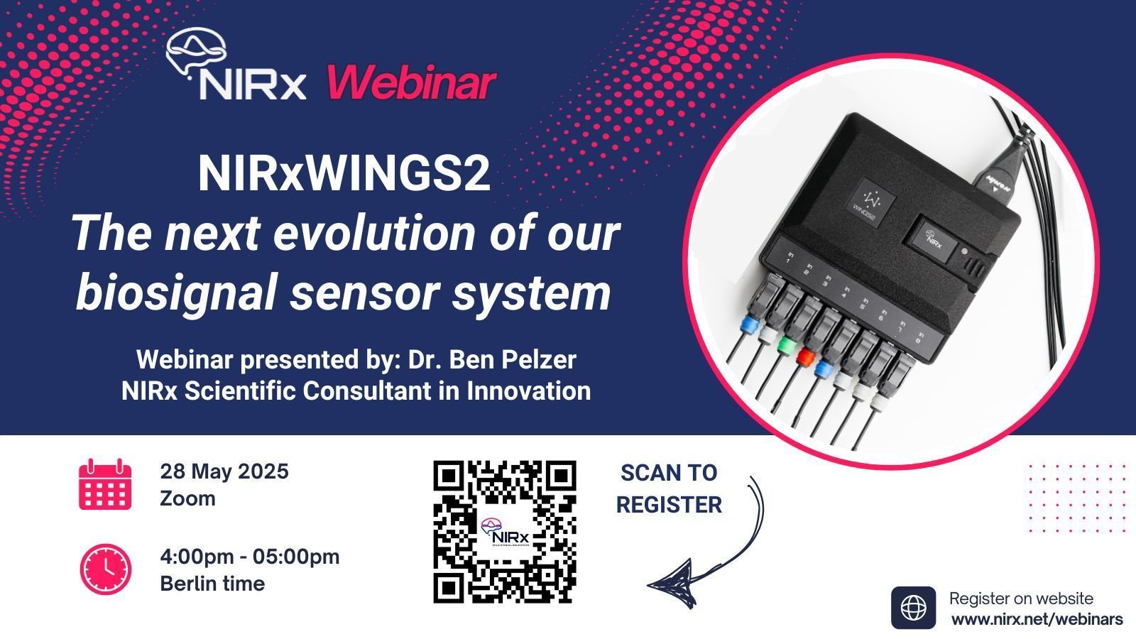
See It in Action – Join Our Webinar
Curious how NIRxWINGS2 can elevate your work? Don’t miss our upcoming live webinar, where we’ll showcase:
How to set up your custom configuration with NIRx BioLink
Strategies to improve data quality across modalities
Real use cases from our global research community
Live Q&A with our NIRx expert support team!
📅 Date: May 28th, 2025
⏰ Time: 4-5 pm, GMT + 2
🔗 Register via Zoom

New Aurora with advanced multiplexing for high-density fNIRS
by Dr. Alexander von Lühmann
What is High-Density fNIRS and Diffuse-Optical Tomography, and why should we care?
In recent years, we have witnessed a comeback of High-density fNIRS studies. Dr. David Boas discusses the recent advances and perspectives in this 2 part-webinar.
HD-fNIRS can improve spatial resolution, depth, and lateral specificity, and can enable image reconstruction of cerebral activation (Diffuse Optical Tomography, DOT).
DOT fNIRS research was initially only done by a small number of experts in the field because it requires special infrastructure, such as many channels, high dynamic range instrumentation, and photon migration simulation algorithms.
DOT fNIRS systems have become more accessible and open toolboxes make it much easier to perform image reconstruction of the data. Here’s a walkthrough of the NIRStoolbox by Dr. Theodore Huppert.
"What are the trade-offs to achieve high-density measurements?"
Excellent signal quality and a good sampling rate are basic requirements for fNIRS research, regardless of conventional or high-density measurements. To achieve high-quality recordings, a trade-off between several challenges has to be addressed:
A high dynamic range is needed to measure channels at short and long source-detector distances. Long channels need brighter sources to see good signal, but brighter sources can saturate short channels, reducing signal quality.
With multiple sources closely packed, avoiding crosstalk is a must. Pure time-multiplexing (turning one source on at a time) in a high-density setup reduces the sampling rate.
The new Aurora: Multi-Level Illumination and Extended Frequency Encoding
We solved these issues above by unlocking two powerful new features of the NIRSport2 with Aurora: Multi-Level Illumination (MLI) and Extended Frequency Encoding (EFE).
Multi-Level Illumination now enables each source to be used multiple times with different intensities in the same measurement cycle.
This way, optimal signal quality can be achieved despite differences in source-detector distances between channels! The result: optical signal quality at an even higher dynamic range, locally optimized.

Diagram showcasing one use case of the MLI feature. If a source has channels at short distances (e.g. <20 mm short separation channels) and at long distances (e.g. usual >30 mm channels), detectors that are too close to a light source might be “blinded” by the amount of light needed to reach the longer channels. By splitting the source illumination sequence into two parts with different light levels, it is possible to first: (A) use lower-powered light to optimally reach detectors at shorter distances; and then (B) use higher-powered light to optimally reach detectors at longer distances.
Extended Frequency Encoding increases the number of simultaneously used frequencies for signal (de)modulation from two to eight.
What does this mean? More sources close by one another can be turned on at the same time without affecting signal quality by creating crosstalk. This saves precious time switching steps and therefore doubles the available sample rate.
Smart Optimization feature: Both MLI and EFE do not have to be configured by the user.
If enabled, our new Smart Optimization feature automatically figures out the best signal quality and sample rate that can be achieved with your montage.
In summary: Your NIRSport2 gets an upgrade for high-density and higher sample rates, both without making any compromises on the outstanding signal quality that the system is known for. We further empower you to perform DOT fNIRS-based tomographic image reconstruction within our NIRSport2 platform.
Get in touch with us at consulting@nirx.net to find out more
New Software Updates Aurora + NIRSite
We are delighted to announce new software versions for Aurora and NIRSite by NIRx. They may already be downloaded from our Help Center!
We are delighted to announce new software versions for Aurora and NIRSite by NIRx. They may already be downloaded from our Help Center! These updates include compatibility for MacOS and the Hyperscan application. Thank you to our team in Berlin for making this happen.

Aurora 2021.4: Data acquisition with NIRSport2
New features and improvements
Support of single device split Hyperscanning in Hyperscan application. Ability to choose two different (max. 8x8) configurations to run from one NSP2 device, support for short channels and accelerometer. Independent visualization of data, separate storage of data.
LSL stream update: Inclusion of subject information (name, age, etc.) in LSL stream metadata.
Full accelerometer data in LSL stream.
Full accelerometer data (100 Hz) in SNIRF file.
Accelerometer samples (in LSL stream, *.acc, and SNIRF file) are corrected for drift of clock between NSP2 and accelerometer.
More default montages & configurations included when Aurora is initially installed.
Bug fixes and changes
Better error messages/handling when writing access to Documents folder is denied by Windows
Validation of "Data root directory" and "Configuration directory" on the settings page (i.e. errors when the directory does not exist or Aurora can't write to it)
Hyperscan data now saved in the Hyperscan folder instead of the Data folder.
Better validation of custom channel names/labels

NIRSite 2021.4: Montage Creation
Bug fixes and changes
Compatibility now spans from macOS Mojave to Big Sur.
Optode positions created in the fNIRS Optodes’ Location Decider (fOLD) can be loaded directly into NIRSite (if saved in ASCII format).
Digpts.txt file is automatically generated in the montage folder.

Additionally, we will be hosting an open webinar 11th May, 10 am EST/ 4 pm CET on Hyperscan: The hyperscanning feature for NIRSport2 + Aurora that will go over many of these features - see you there!
Would you like to learn more? Feel free to reach out to our support team. We’d be happy to hear from you.

Upcoming Webinars
Including a guest lecture by Dr. Ted Huppert
We are pleased to announce that we have three new webinars coming up! The first upcoming webinar will be a guest webinar by Dr. Ted Huppert (University of Pittsburgh). A week later the second webinar will focus on fNIRS functional connectivity analysis, and finally we will walk through the new Aurora fNIRS software. See below for more details, exact dates and registration links!

Upcoming Webinars
Co-registering fNIRS data & structural MRI data in MATLAB Brain AnalyzIR Toolbox (NIRS Toolbox) - March 28th 2019

We are honored to have Dr. Ted Huppert (University of Pittsburgh, developer of NIRS Toolbox) join us as guest speaker for a special NIRx webinar focusing on co-registering fNIRS data & structural MRI data in the Brain AnalyzIR Toolbox.
During the Webinar, Dr. Huppert will go over the import of anatomical (MRI) information, probe (montage) registration to anatomical landmarks (accounting for head size variations), optical forward model simulations, image reconstruction basics, and anatomically defined region-of-interest methods. If you are curious to learn more about the work of Dr. Huppert, visit his personal website http://huppertlab.net/, or view a recording of one of his lectures here: https://nirx.net/fnirs-analysis.
The Co-Registering fNIRS/MRI in NIRS Toolbox webinar is scheduled for:
March 28, 3PM CET, 10am EDT.
Studying functional connectivity with fNIRS - April 3rd 2019

How do different ares of the brain interact when the mind is at rest? Is there a causal effect between activation to a cognitive task in one area and simultaneous activity in another? Are there functional networks hidden behind our data?
For over a decade, functional connectivity has been receiving increased attention and curiosity of many neuroscientists. Several neuroimaging modalities can be applied to better understand functional networks, but the exquisite temporal resolution of fNIRS offers a perfect opportunity for a closer look at these networks.
In the “Studying functional connectivity with fNIRS” webinar, we will review concepts of functional connectivity and provide a simple demonstration of how to run a connectivity analysis with fNIRS data using the Brain AnalyzIR Toolbox.
The Studying functional connectivity with fNIRS webinar is scheduled for:
April 3, 4pm CET, 10am EDT.
Visit our online Support Center to register for this Webinar!
Overview of Aurora fNIRS
The brand-new Aurora fNIRS software package is intuitive, easy-to-use, and compatible with multiple operating systems. Developed to take a whole new approach to user instrument control and data acquisition, Aurora fNIRS introduces many new features as well as a fresh and elegant new look.

Among many added features, Aurora fNIRS comes with new optimized signal diagnostics to ensure that signal quality is optimal for every measurement. When data is recorded, the user has quick access to high-end whole-head 3d data visualization. And with the integrated LSL protocol, Aurora fNIRS is ready for real-time data analysis and multi-modal measurements.
Interested to learn all new advantages, and see how Aurora fNIRS differs from NIRStar?
The Overview of Aurora fNIRS webinar is scheduled for:
April 11, 4pm CET, 10am EDT.
Learn more about this webinar, as well as others, by visiting our Webinars page.
NIRSite 2.0
We are excited to announce NIRSite 2.0!
The second installment of NIRSite has arrived! Read about the new features here, such as easy addition of short-distance channels, automatic export of montages, renaming source and detector labels, and much more!

Following the success of the first release of NIRSite, we are excited to share with you the updated and much improved NIRSite 2.0. Packed with new features and important bug fixes, NIRSite 2.0 makes creation of montages / optode-arrangements easier than ever!

What’s new in NIRSite 2.0?
Effortless addition of short-distance channels
Manual manipulation of measurement channels
Renaming source and detector labels
Creation of montages in the two-dimensional view
Automatic export to NIRStar and Aurora montage folders
Visit the NIRx Help Center to learn more about NIRSite 2.0, and download your own copy free of charge.
In addition, we warmly invite you to attend the upcoming NIRSite 2.0 Webinar, scheduled for March 7 2019. Visit our Webinar page for more details!